OCAD MSK
History
History of dislocations

Discussion
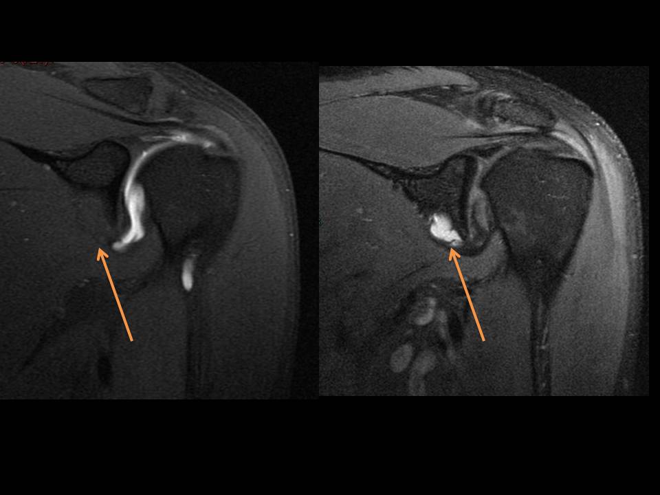
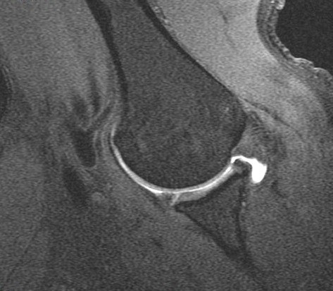
32 y/o s/p multiple dislocations. First image is of pre injection axial PD fat sat and post injection T1. The cyst is clearly visible on the pre image. No communication with the joint. Coronal images same circumstances as the axials. The ABER image demonstrates a tear of the anterior inferior labrum (this was felt to be a Perthes lesion).
Diagnosis
Non communicating labral cyst