OCAD MSK
History
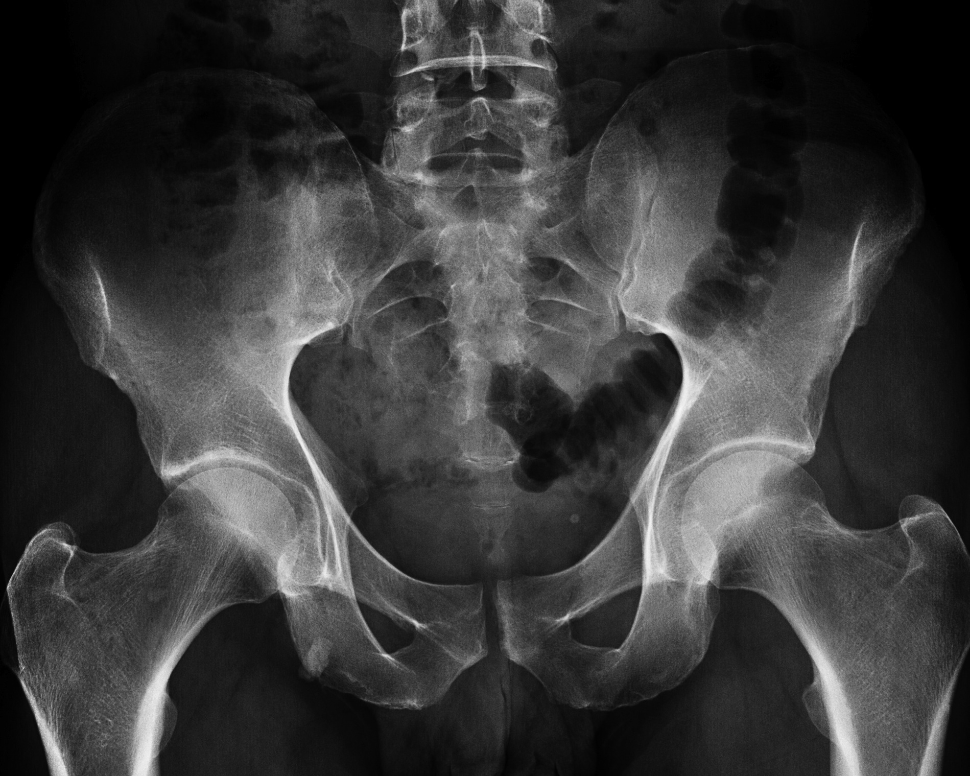
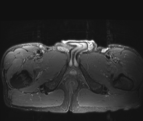
Jogger with pain
Discussion
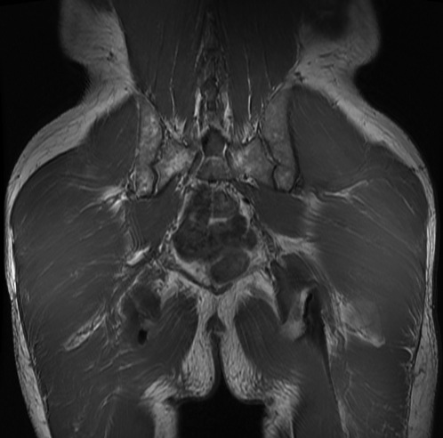
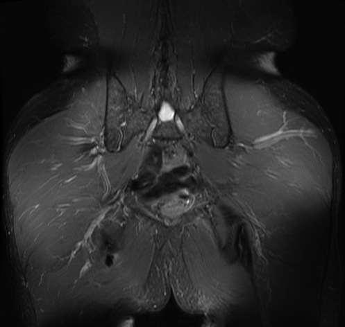
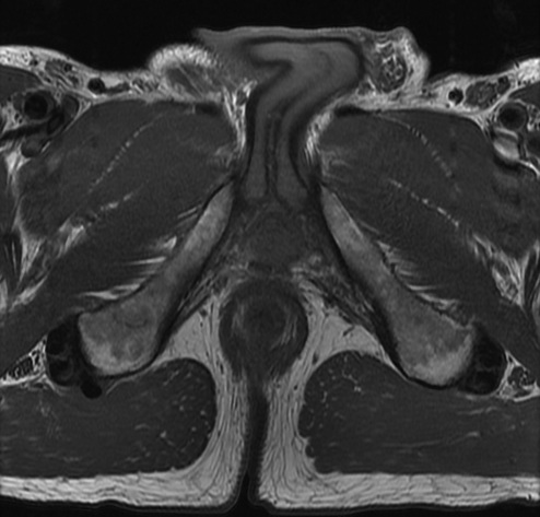
AP pelvis shows a sclerotic density in the right ischium, but a small component extends past the cortex, suggesting it is external to the bone. MRI shows a likely extruded calcific tendonitis of the right hamstring origin. Reference article.
Diagnosis
Calcific tendonitis hamstring