OCAD MSK
History
Discussion
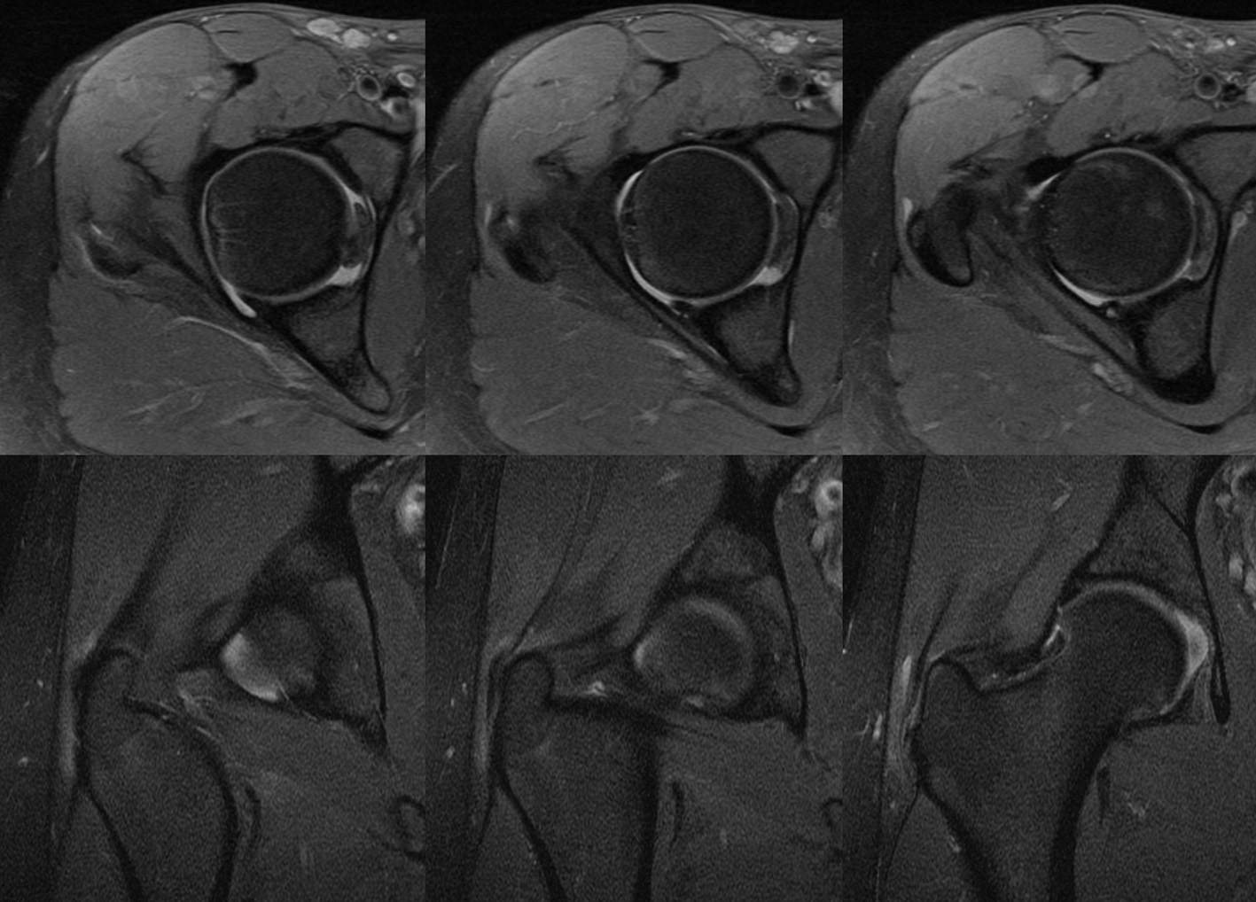
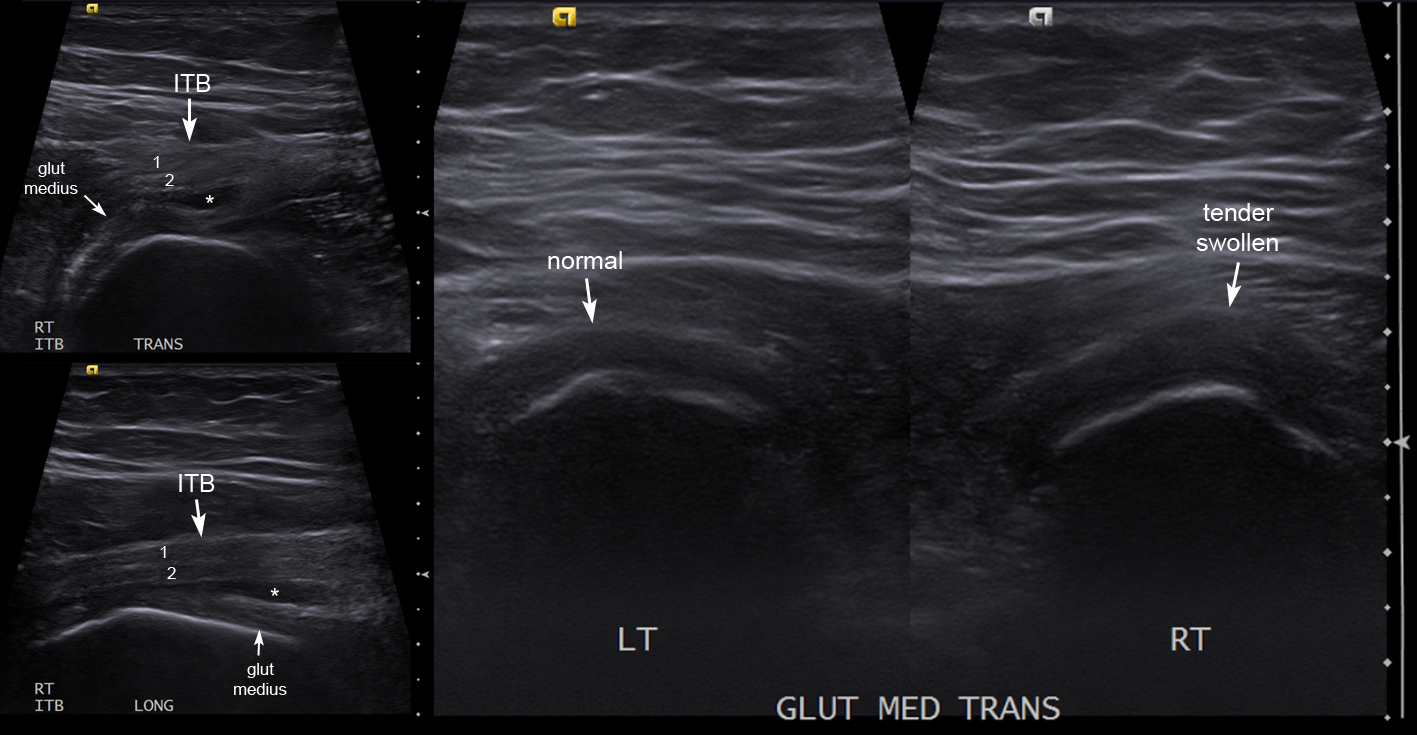
MRI shows subtle but definite hyperintense thickening of the gluteus medius tendon. US showed no obvious difference in tendon echotexture, but abnormal right gluteus medius tendon was tender and diffusely swollen. Also small amount of fluid in sub-maximus/sub-ITB trochanteric bursa (*). Note the normal 'bi-laminar' appearance of the ITB along its posterior half at greater trochanter level due to contributions from gluteus maximus and TFL (layers 1 and 2) --> this is routinely visualised with US but is difficult to appreciate on MRI without an understanding gluteus maximus tendon anatomy.
Diagnosis
gluteus medius tendonitis