OCAD MSK
History
Slowly growing subcutaneous mass
Discussion
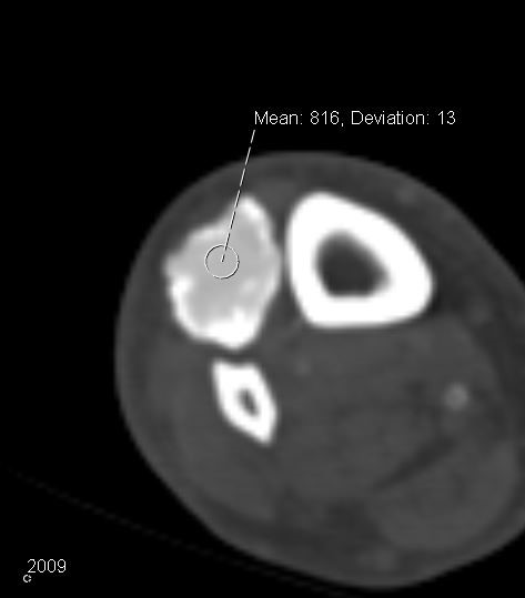
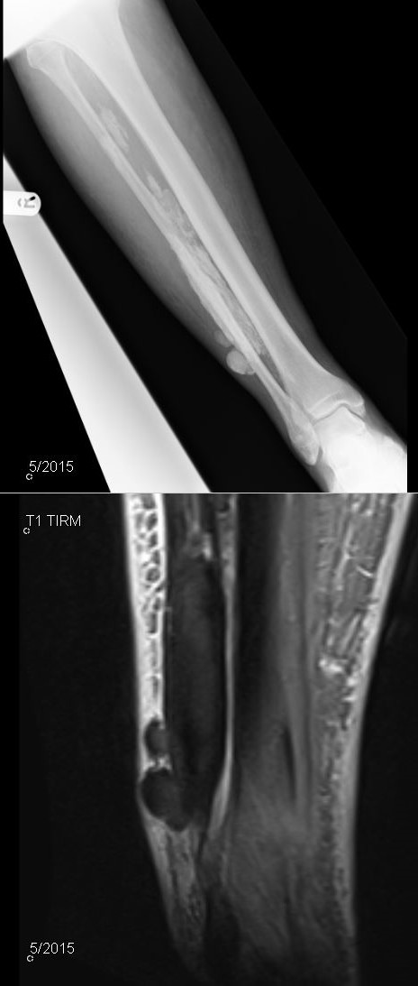
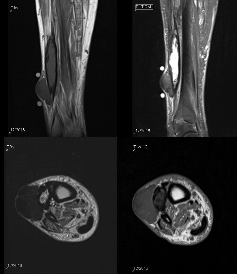
70 yo/m with increasing lump over the anterolateral tibia. Images are from 2009, 5/2015 and 12/2016.
The large mass in the ant. compartment is stable in size, now with central liquefaction. Adjacent is slightly growing subcutaneous mass, also showing calcifications. It seems to be originating from the larger mass, with herniation through the fascia. Reference article.
Diagnosis
Calcific myonecrosis