OCAD MSK
History
28F with knee pain and locking
Discussion
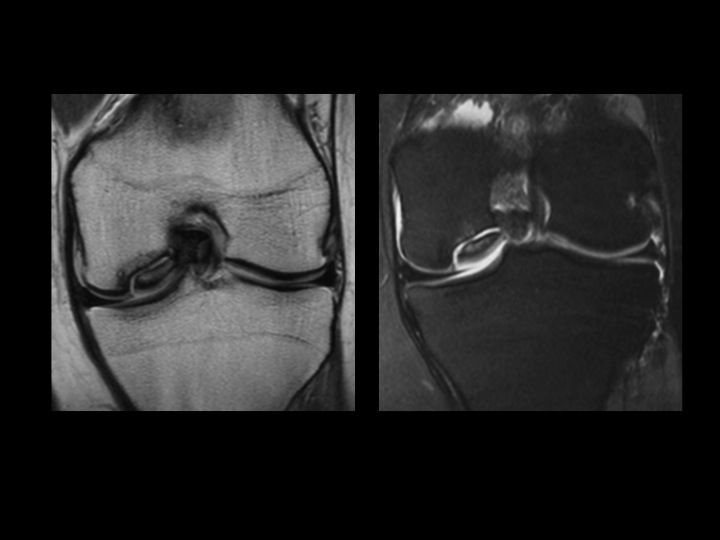
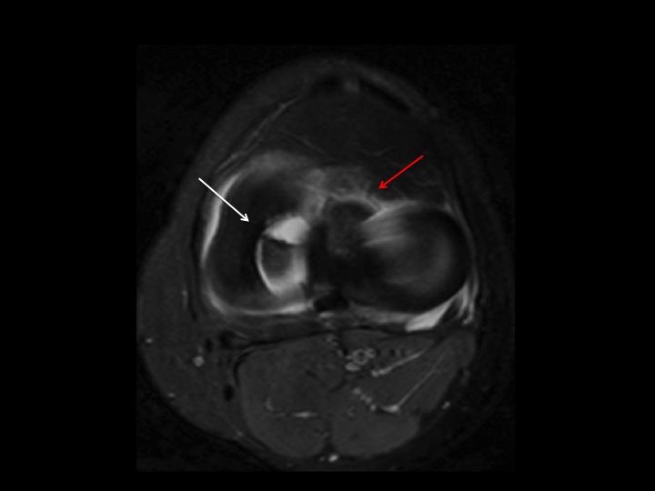
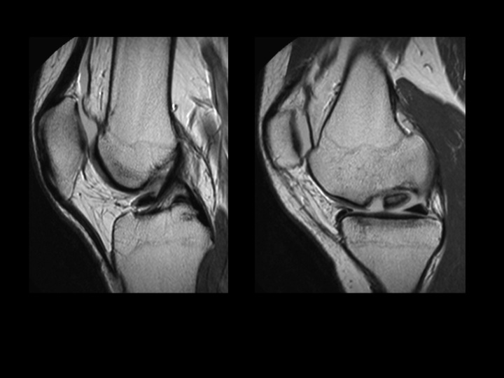
No classification is perfect, but I generally use Andersons MRI classification for OCLs, regardless the affected bone or joint. In the Cor images this looks like a straightfoward Type III OCL, but the axial image shows the anterior margin of the OC fragment is truncated (white arrow) and displaced into the anterior intercondylar notch (red arrow). This is clearly seen in the Sag PD images. I didnt quite know how to classify this? Upgrade to Type IV? 3 1/2? Of course its all unstable and I described it as such, but no classification system is perfect.
Diagnosis
unstable osteochondral lesion